|
|
Infected Bulla
General Considerations
- A bulla is characteristically thin-walled -- < 1 mm
- Air-filled space
- Contained within the lung
- 1 cm in size or greater when distended
- Walls may be formed by pleura, septa, or compressed lung tissue
Clinical Findings
- Patients with infected bullae tend to be less sick than those with a lung abscess
- Can be asymptomatic
- Cough
- Purulent sputum
- Elevated white blood count
Imaging Findings
- Seen more in upper lobes
- Thin-walled, sharply demarcated areas containing no visible blood vessels on conventional radiography
- Only portion of wall is usually seen on conventional radiography
- They tend to trap air
- May become larger on expiration
- Signs of an infected bulla
- Air-fluid level is best sign
- Prior existence of a non-infected bulla in same region helps diagnosis
- Differentiation from lung abscess
- Bulla contains less fluid
- Much thinner wall
- No surrounding pneumonitis
- Patients less sick with infected bulla
Differential Diagnosis
- Other air-containing pulmonary structures
Other Air-containing Structures in the Lung |
Name |
Remarks |
Pneumatocoele |
Thin-walled (< 1mm), gas-filled space in the lung developing in association with acute pneumonia, such as staph, and frequently transient |
Cavity |
Gas-containing space in the lung having a wall > 1 mm thick |
Cyst |
Thin-walled, air- or fluid-filled, with a wall that contains respiratory epithelium, cartilage, smooth muscle and glands |
Bleb |
Intrapleural cystic space, typically very tiny |
Treatment
- Clearing may take weeks to months
- Antibiotics are usually administered
- Bronchoscopy is frequently not helpful
Complications
- Enlarge progressively over a period of months to years
- Bullae may become so large as to render the remaining normal lung almost invisible, pancaked atop the hemidiaphragm = vanishing lung syndrome

- Most are associated with emphysema
- May lead to pneumothorax
 
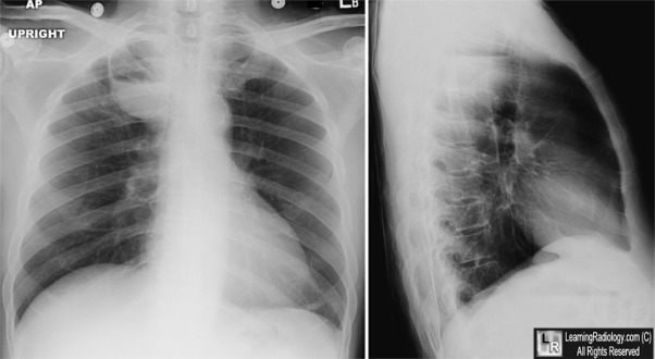
Infected Bulla. Image on left shows a thin-walled bulla in the right upper lobe (white arrow) several months before photos on right which demonstrate an air0fluid level in the same bulla on the frontal (white arrow) and lateral (black arrow) views.
For more information, click on the link if you see this icon 
For this same photo without the annotations, click here or here
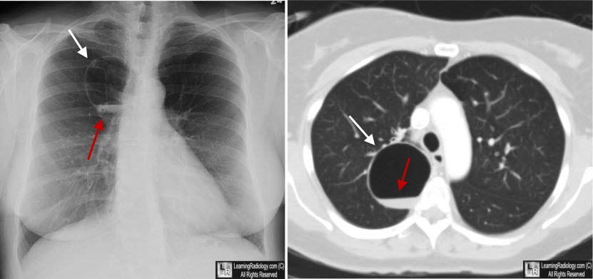
Infected Bulla. Image on left shows a thin-walled bulla in the right upper lobe (white arrow) with an air-fluid level (red arrow). CT of the chest at the same time again demonstrates an air-fluid level (red arrow) in the bulla (white arrow).
Chest. 2006;130:1942-1946. “A 57-Year-Old Man With a Fluid-Containing Lung Cavity” Chandra, D; Soubra, S and Musher, D.
|
|
|
